
Notes on Anatomy and Physiology The Vertebrae
Dura Mater Fat in Epidural Space Iliolumbar Ligament Inferior Articular Process Intertransverse Ligaments Intervertebral Discs L1 (1st Lumbar Vertebra) L2 (2nd Lumbar Vertebra) L3 (3rd Lumbar Vertebra) L4 (4th Lumbar Vertebra) L5 (5th Lumbar Vertebra) Nucleus Pulposus Pia Mater Posterior Sacroiliac Ligament Radiate Ligaments Sacroiliac Joint
Thoracic vertebrae anatomy, function & thoracic vertebrae injury
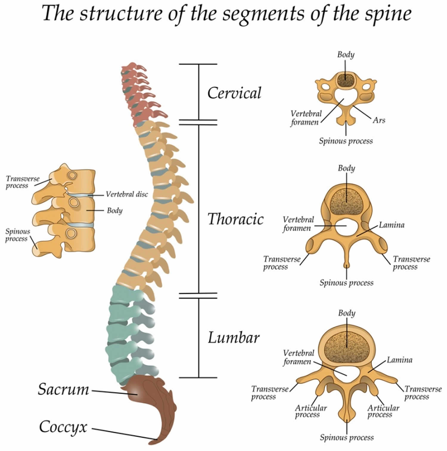
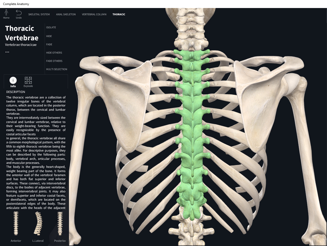
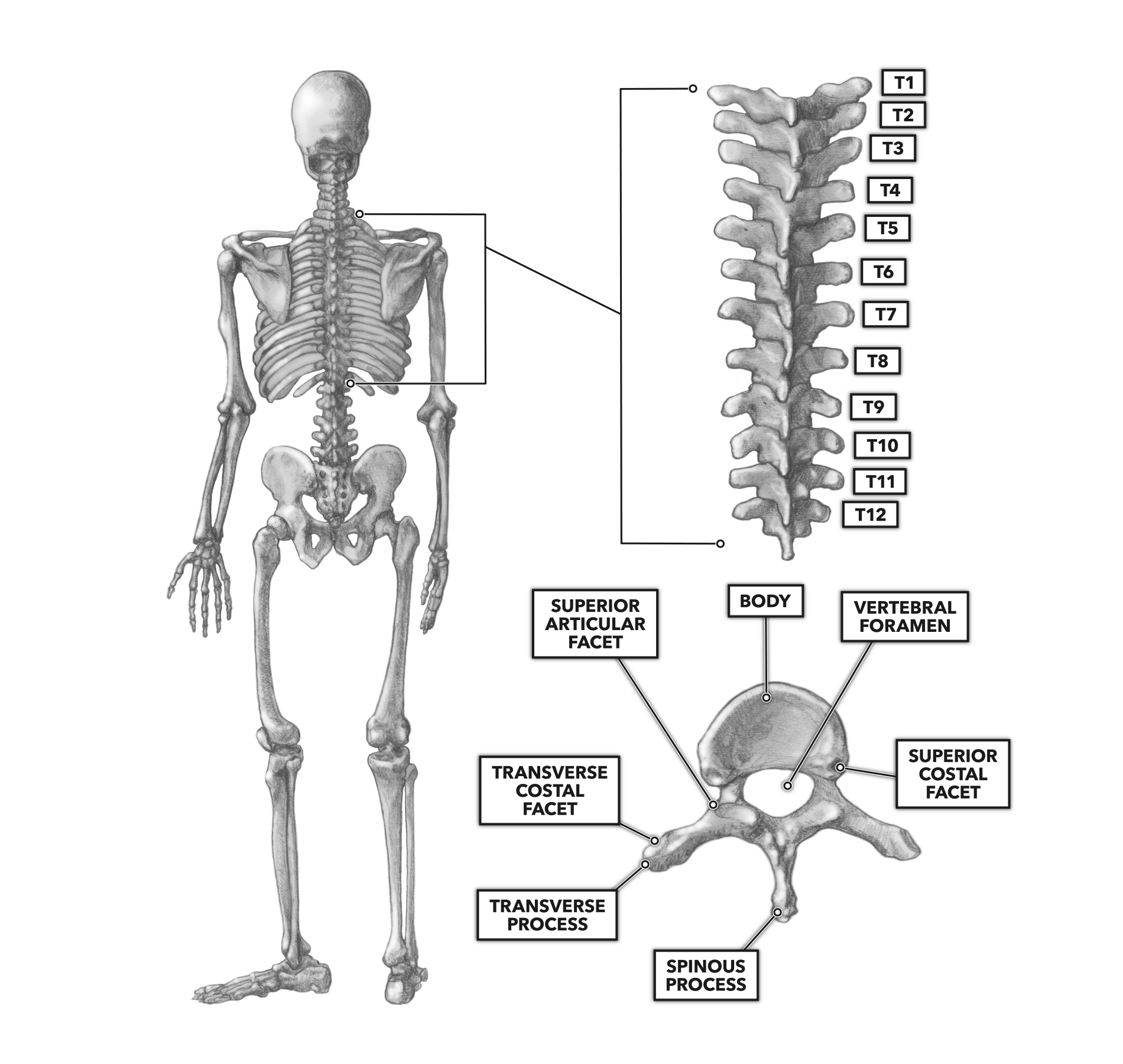
Published on May 24th 2022 by staff What is Thoracic Spine The thoracic spine is the second and longest part of the spinal column, consisting of 12 lumbar vertebrae, T1-T12. These 12 bones are separated from each other by intervertebral discs. Their primary role is to form the thoracic cage that protects the heart, lungs, and esophagus.
Thoracic vertebrae diagram
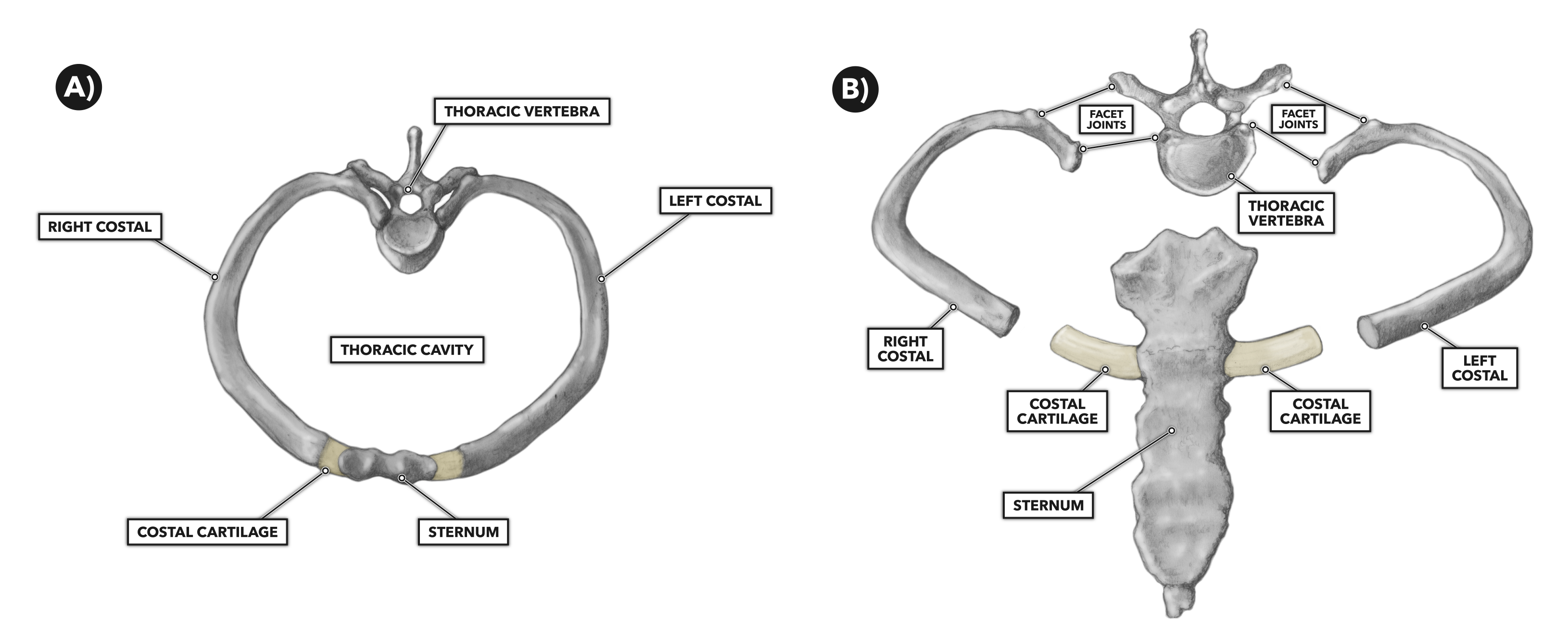
Moving forward with the skeletal scaffold of the thorax, we have the thoracic skeleton.It is made up of the sternum, twelve pairs of ribs, twelve thoracic vertebrae, and interconnecting joints.The main thoracic joints include the intervertebral discs, costovertebral, sternocostal, sternoclavicular, costochondral, and interchondral joints.. Running between every two adjacent ribs are anatomical.
Thoracic vertebrae structure Science online
The thoracic spine is the second segment of the vertebral column, located between the cervical and lumbar vertebral segments.It consists of twelve vertebrae, which are separated by intervertebral discs.. Along with the sternum and ribs, the thoracic spine forms part of the thoracic cage.This bony structure helps protect the internal viscera - such as the heart, lungs and oesophagus.
Third Thoracic Vertebra Viewed from Above ClipArt ETC
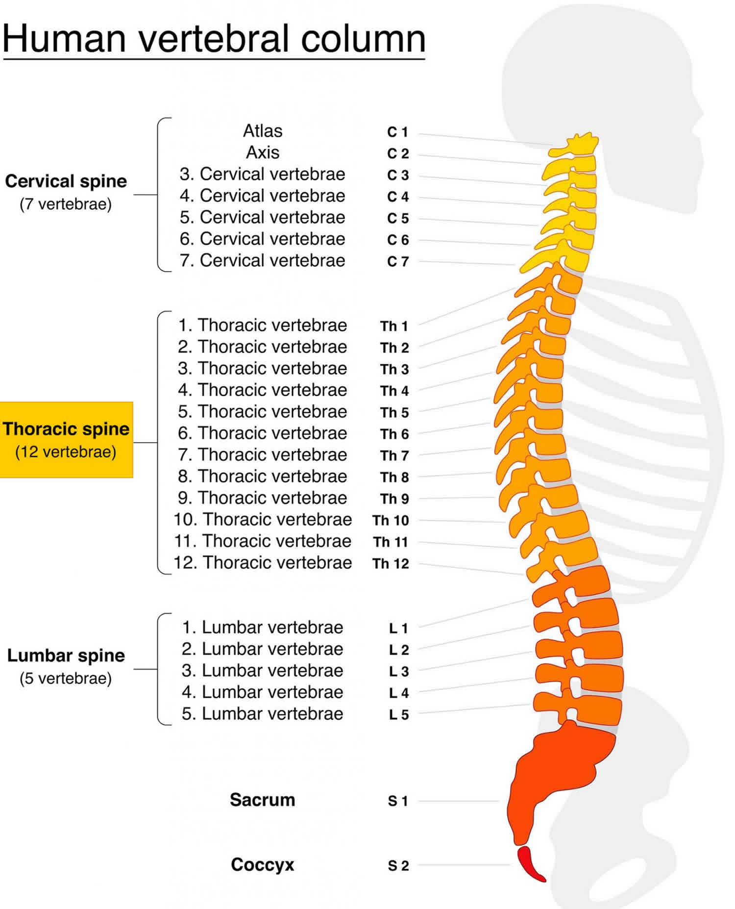
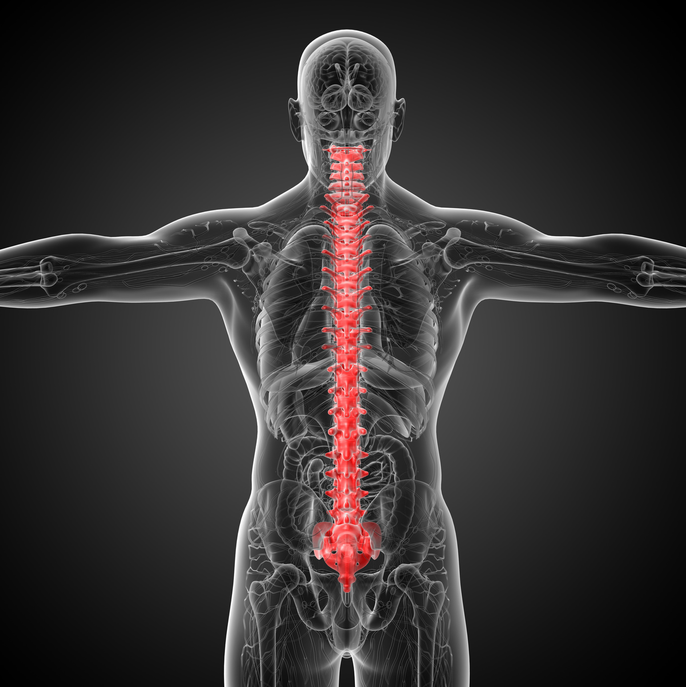
Anatomy Explorer Annulus Fibrosus Anterior Longitudinal Ligament Anterior Sacroiliac Ligament Aorta Arachnoid Mater C1 (Atlas) - 1st Cervical Vertebra C2 (Axis) - 2nd Cervical Vertebra C3 (3rd Cervical Vertebra) C4 (4th Cervical Vertebra) C5 (5th Cervical Vertebra) C6 (6th Cervical Vertebra) C7 (7th Cervical Vertebra) Coccyx
Anatomy Of Thoracic Vertebrae
Anatomy Explorer Annulus Fibrosus Anterior Longitudinal Ligament Anterior Sacroiliac Ligament Aorta Arachnoid Mater C1 (Atlas) - 1st Cervical Vertebra C2 (Axis) - 2nd Cervical Vertebra C3 (3rd Cervical Vertebra) C4 (4th Cervical Vertebra) C5 (5th Cervical Vertebra) C6 (6th Cervical Vertebra) C7 (7th Cervical Vertebra) Coccyx
Thoracic Vertebra Photograph by Asklepios Medical Atlas Pixels
Causes of Upper Back Pain Video The Thoracic Spine: Roles and Functions The thoracic spine is one of the four major regions of the spine. The thoracic spine has 12 vertebrae stacked on top of each other, labeled from T1 down to T12.
Thoracic Vertebrae Anatomy and Pathology Kenhub
The thoracic spinal vertebrae consist of 12 total vertebrae and are located between the cervical vertebrae (which begin at the base of the skull) and the lumbar spinal vertebrae. The.
Bones Vertebral Column, Thoracic Region. Anatomy & Physiology
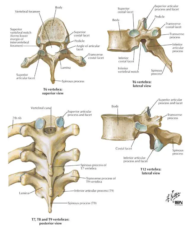
Anatomy. The T7 vertebra is located in the middle of the thoracic spinal column inferior to the T6 vertebra and superior to the T8 vertebra.mycontentbreak. The anterior portion of the T7 vertebra is made of a wide, heart-shaped cylinder of bone known as the centrum or vertebral body. The centrum provides most of the strength to the vertebra and.

Thoracic Vertebrae
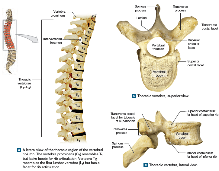
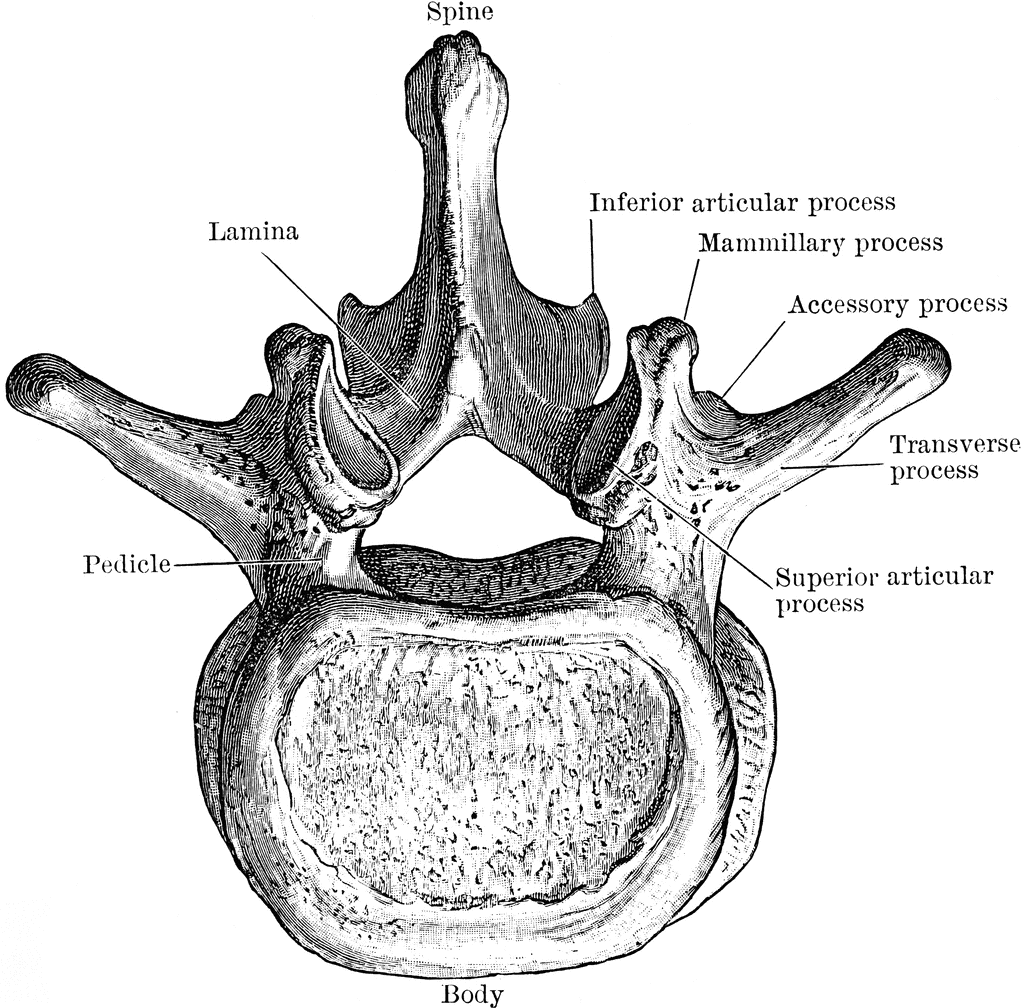
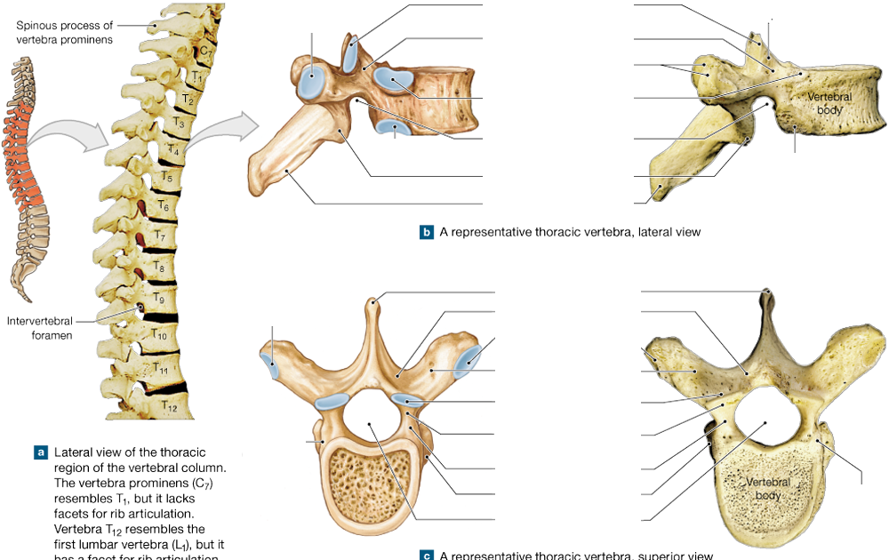
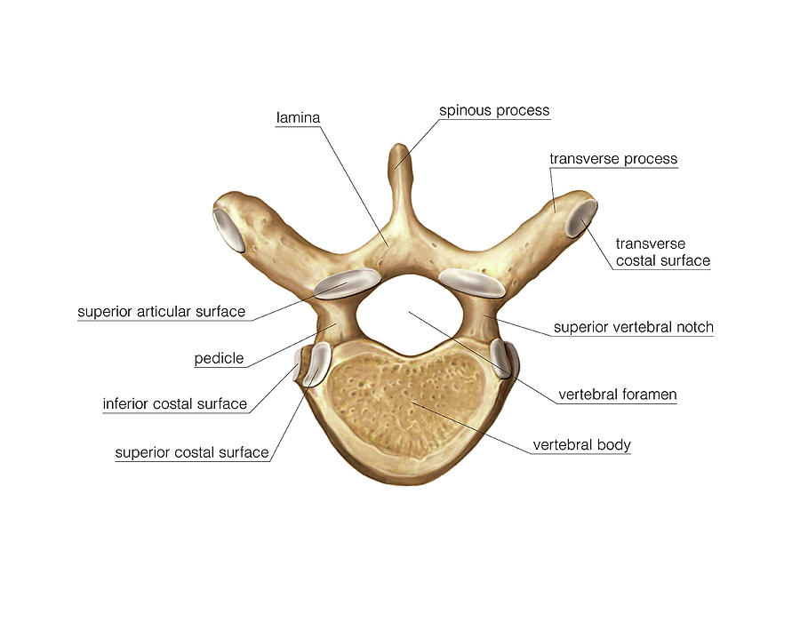
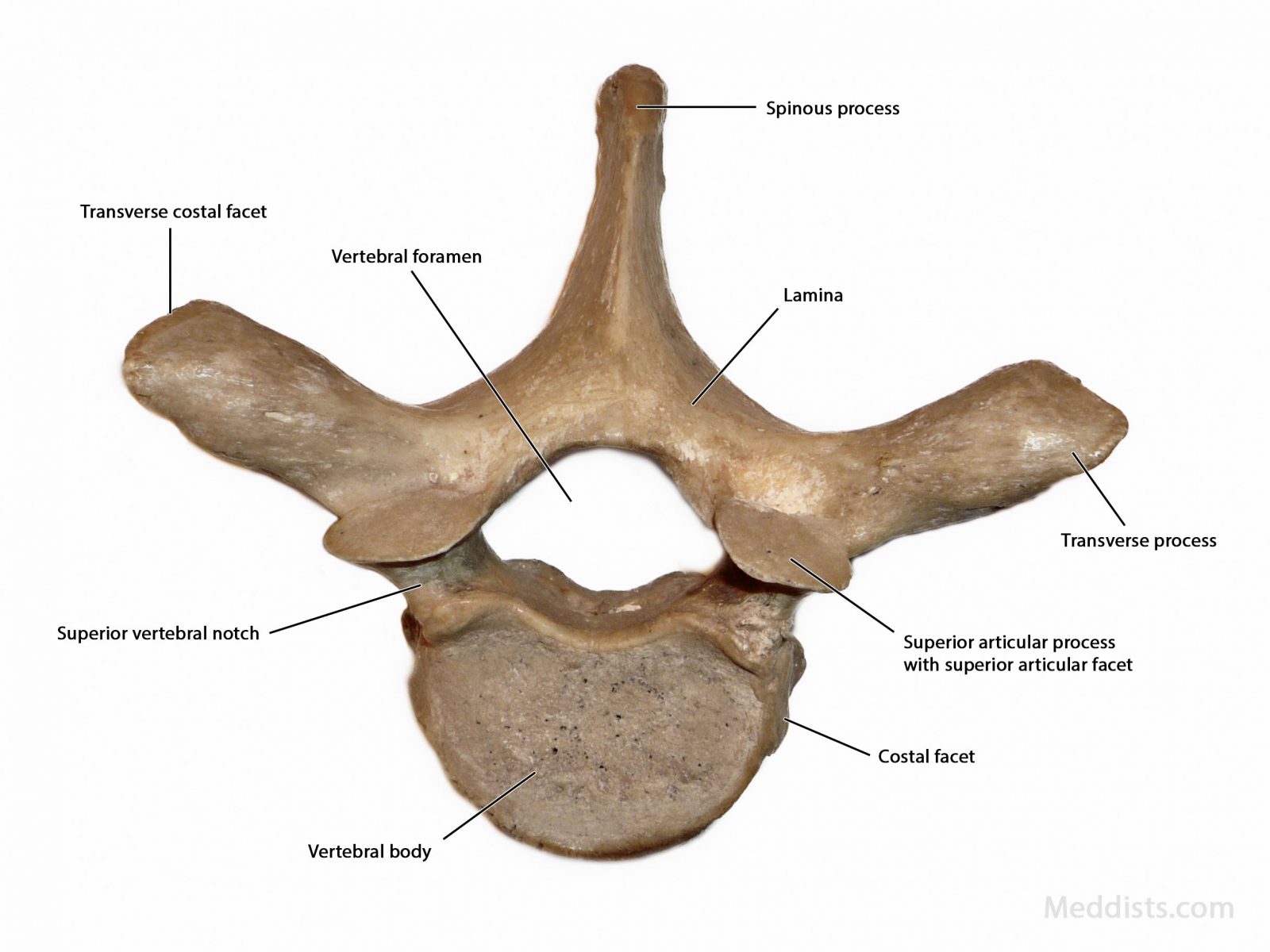
Thoracic vertebrae T2 to T8 are all similar, although they do gradually get bigger while going down the spine. A typical thoracic vertebra consists of the following: Vertebral body. This thick, bony front of the vertebra is a rounded heart shape (as viewed from above) in the thoracic spine region. The vertebral bodies stack on top of each other.
CrossFit The Thoracic Vertebrae and Other Thoracic Bones
1/2 Synonyms: Vertebrae T1-T12 The twelve thoracic vertebrae are strong bones that are located in the middle of the vertebral column, sandwhiched between the cervical ones above and the lumbar vertebrae below. Like typical vertebrae, they are separated by intervertebral discs.
Anatomy of the Thorax → Thoracic Vertebral Column
Anatomy of the Thoracic Spine. In technical terms, your spinal column at the mid and upper back levels is called the thoracic spine. The thoracic spine is comprised of 12 spinal bones connected to and occupying the same level in your body as your 12 ribs. In fact, the thoracic spine works with the ribs to create a protected space—your rib.
Grassy Knoll JFK Autopsy Photos www.GrassyKnoll.US
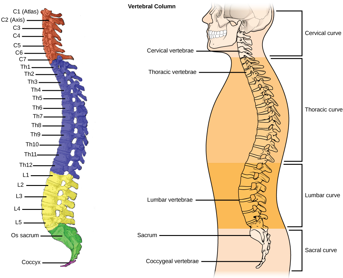
The vertebral column has four main functions: Protection - encloses and protects the spinal cord within the spinal canal. Support - carries the weight of the body above the pelvis. Axis - forms the central axis of the body. Movement - has roles in both posture and movement. Fig 1 - The vertebral column viewed from the side.
CrossFit The Thoracic Vertebrae
Pia Mater Posterior Sacroiliac Ligament Radiate Ligaments Sacroiliac Joint Sacrospinous Ligament Sacrotuberous Ligament Sacrum Spinal Ganglion Spinous Process
Thoracic vertebrae anatomy, function & thoracic vertebrae injury
The T12 vertebra is the twelfth thoracic vertebra in the spine of the human body. It is part of the spinal column, which supports the top of the human body. The spinal column extends.
Thoracic Vertebrae Brain and Spinal Cord
The tenth thoracic vertebra (T10) is one of twelve vertebrae that make up the central section of the vertebral column. The spine consists of three vertebral columns, including the cervical.